Imaging Could Take the Place of Repeat Prostate Cancer Biopsies

One of the most significant areas of success in modern medicine is prostate cancer treatment and prevention.
For starters, the five-year relative survival rate—the percentage of people who live at least five additional years following their prostate cancer diagnosis—is 96.8 percent. That's far better than the 68 percent rate in 1975 and includes all stages combined. When detected in the localized stage, prostate cancer is almost 100 percent survivable.
As the medical community learns more, it's not just the duration of those lives but also the quality that has improved. For instance, better precision in diagnosing low-risk versus high-risk cancer has helped more men go on active surveillance—simply monitoring the patient's prostate cancer—instead of having potentially unnecessary surgery that can have lifelong side effects. Biopsy protocols in testing for prostate cancer, especially for people with low-grade cancer who are on active surveillance, have improved greatly as well.
But what if there were some way to reduce the number and frequency of invasive biopsies that patients on active surveillance need? With cutting-edge advances in imaging becoming more accessible, many professionals in the healthcare community think that's an area worth exploring.
Active surveillance and ongoing testing
As recently as 10 years ago, a majority of people diagnosed with even low-grade, low-risk prostate cancer were treated immediately with surgery or radiation. Today, active surveillance is another option for many patients.
Candidates for active surveillance must meet certain benchmarks, such as having cancer confined to the prostate, having prostate-specific antigen (PSA) values of 10 nanograms per milliliter (ng/mL) or less, and a Gleason score of 6 or less, according to the National Cancer Institute.
A confirmation biopsy six months after the initial diagnosis that establishes the cancer as low grade is usually the next step.
"Active surveillance was a boon for many patients with low-risk cancers," said Murugesan Manoharan, M.D., the chief of urologic oncologic surgery at the Baptist Health Medical Group in South Florida. "But when I have a Gleason score that shows a low-risk prostate cancer, we will always do another biopsy in another six months to make sure we're not missing anything."
Until now, repeat biopsies every year had been the standard for a lot of practitioners. Those were in addition to more frequent PSA tests and digital rectal exams. Advanced imaging may mean that window can be pushed out to two years—maybe even three—between biopsies. All thanks to tesla (not that one).
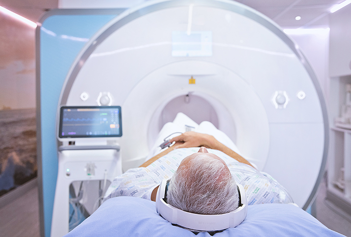
MRI scans spare patients biopsies
In this case, we're talking about tesla as a unit of measurement used to express magnetic flux density. Magnetism is the key component of MRI, which stands for magnetic resonance imaging.
When it comes to imaging that can indirectly determine whether a person's prostate cancer is stable and remains low-risk without a biopsy, the technology should be the best available.
"With MRIs, not every one of them is equal. Some are more equal than others," Manoharan said. "The magnet of the MRI is the most important thing, and that's measured in the unit tesla. What we need is to make sure we have a three-tesla [3-T] magnet when we do a prostate MRI, and we have to do them in a particular sequence."
This is what is known as 3-T multiparametric magnetic resonance imaging, and studies show it's very good at predicting cancer reclassification among active surveillance patients. In this instance, reclassification means clinicians recognize if the cancer has changed.
The usefulness of a tool, though, is dependent on the skill of the person wielding it.
"The other thing is you need a very good radiologist dedicated to MRIs to read it," Manoharan explained. "The good thing about a 3-T MRI is that it may miss a very low-risk cancer but it rarely misses a significant cancer. So if a patient is due for a biopsy, but his PSA and everything is good and the MRI shows that there are no significant lesions, it puts my mind at rest that even if I'm missing something, it's only low-risk cancer."
There's always a backup plan
Healthcare providers helping a person navigate something as serious as a cancer diagnosis always have a backup plan.
In the case of using 3-T MRI to help avoid frequent repeat biopsies, any sign the cancer has changed is going to trigger a biopsy. Even in that event, however, advanced imaging can help ensure the accuracy of the procedure.
One such system is MRI-ultrasound fusion technology, which uses software to combine MRI and ultrasound scans to help guide the biopsy needle to the correct spot.
"If the MRI shows lesions—every conversation is a bit nuanced—you're going to want to make a decision on performing a biopsy," said Aditya Bagrodia, M.D., a urologic oncologist with University of California San Diego Health. "And then the gold standard would probably be an MRI-ultrasound fusion biopsy. It just makes sure that you're hitting the areas that are likely to be cancer."
Conclusions
Prostate cancer is far less deadly than it was even a few decades ago, and many people are able to live higher-quality, fuller lives because of the advances that have been achieved. Being able to keep a close eye on slow-growing, low-risk prostate cancer while limiting invasive procedures as much as possible is a big part of that progress.
Cutting-edge imaging techniques incorporated into active surveillance protocols help immensely.
"You can avoid multiple follow-up biopsies," Manoharan said. "I feel very certain that you need to do one follow-up biopsy, but after that, if you want to avoid it, you can employ the MRI along with other clinical findings. Secondly, if you want to avoid doing it as frequently, you can avoid doing it every year. You can do it [a biopsy] every other year or every three years."


