Here's What You Can Expect at a Testicular Ultrasound

If a doctor has recommended that you get a testicular ultrasound, you may be feeling curious, anxious or even embarrassed about what's next. If so, you'll be happy to learn that getting one is truly no big deal.
Overview of a testicular ultrasound
Ultrasound, or sonography, is a diagnostic test that uses high-frequency waves to generate images of the body's soft-tissue structures, which aren't captured well by X-ray.
A testicular ultrasound, or scrotal ultrasound, produces images of the testicles, their blood vessels and the encompassing scrotum. The procedure is recommended if a patient reports a testicular injury, pain, swelling or a lump.
This form of imaging can be used to detect testicular cancer, illustrate the shape and size of a lump, and distinguish a fluid-filled mass, such as a cyst, from a more concerning solid one. In addition, ultrasound can detect testicular torsion, a painful and serious medical problem that occurs when a testicle rotates and twists the spermatic cord, and other issues with scrotal blood flow. Ultrasound can be used for an array of other testicular problems, including epididymitis, or inflammation of the tube that stores and transports sperm, infertility and undescended testicles.
Ultrasound is an advantageous form of imaging because it requires no needles or injections. While additional imaging, such as computed tomography (CT), magnetic resonance imaging (MRI) or positron emission tomography (PET) scans, may be recommended as a follow-up to provide a more detailed depiction of a mass, ultrasound prevents unnecessary exposure to dangerous radiation. It is also cheaper, quicker and well-tolerated by patients.
What to expect
The ultrasound will be performed by a trained sonographer and later interpreted by your doctor. You will undress from the waist down and put on a hospital gown. The ultrasound technician will explain the procedure and have you lie on your back on the exam table, positioning a towel under your scrotum to elevate it, with another towel on your penis for privacy.
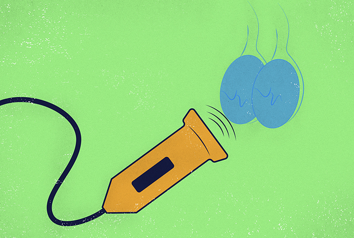
A water-based gel will be applied to the area to ensure good conduction of sound waves and an easy glide for the instrument. An ultrasound transducer emits high-frequency sound waves (inaudible to our ears) and records the echoes that bounce back, indicating the size, shape and depths of the structures beneath, and generating a real-time picture on the monitor.
The sonographer will move the transducer back and forth over the scrotum to take ample images at different angles. The gel may be cold, and you may feel pressure from the transducer. When the procedure is over, there are no restrictions on activities.
Preparation and common myths
There are no limits on eating or drinking beforehand. Medications are rarely a problem, but it's a good idea to double-check with your doctor to ensure you don't need to discontinue any of yours.
While it's commonly believed that you have to shave the groin area before a testicular ultrasound, grooming is entirely up to you. Trimming may be a good idea if you have thick pubic hair, because it can mildly reduce sound penetration from the transducer, but your technician won't mind either way. Another myth is that the process is uncomfortable. This may be true if you're already experiencing tenderness, but if not, the process is painless (albeit a bit chilly, thanks to the gel).
Perhaps the most common misconception about scrotal ultrasounds is that they're awkward. Technicians perform ultrasounds all day long, so your ultrasound is no different from countless others. Typically, techs are skilled at making you feel at ease, and afterward, many guys admit the process was much easier than they had expected. If you're still anxious, an ultrasound typically takes all of 10 to 20 minutes—it will be over before you know it.


