MRIs: A Useful Tool in Detecting Endometriosis and Adenomyosis

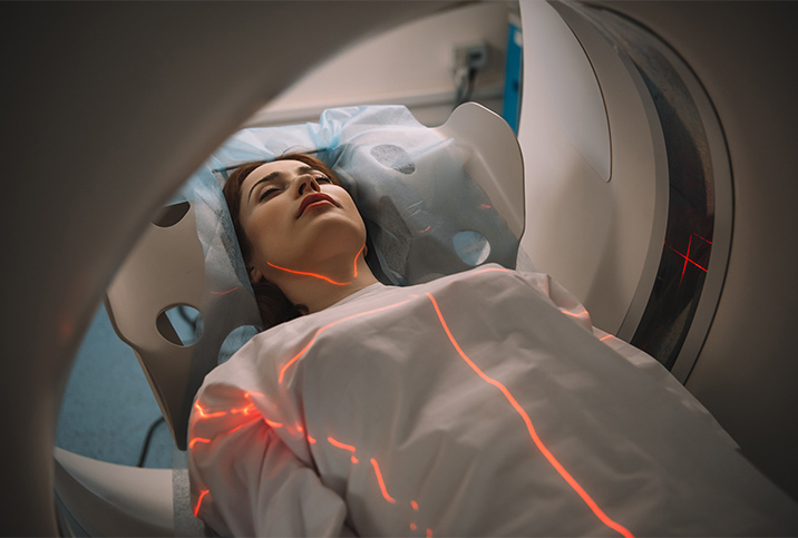
Magnetic resonance imaging (MRI) may be an unexpected gynecological tool, but the technology is well suited for diagnosing endometriosis and adenomyosis in some cases.
As a noninvasive detector of soft tissue beyond what the eye and camera can see and a nonradioactive technology—especially valuable for menopausal women, who are prone to gynecological cancers—MRIs can be the trick to detecting extensive endometriosis and the rarer, but even more invasive, adenomyosis.
However, this junction of radiology and women's health requires uncommon expertise and is far from being the most common route to diagnosing the conditions.
The gold standard for endometriosis diagnosis is laparoscopic imaging, according to Tara Scott, M.D., an OB-GYN in Akron, Ohio.
"The only official way to make the diagnosis of endometriosis is with diagnostic laparoscopy to visualize implants directly. If an endometrioma is found—a chocolate cyst or an ovarian cyst full of endometriosis—this can be visualized on ultrasound. MRI can see different indications of changes of pelvic tissues," Scott explained.
It should be noted that a chocolate cyst is nowhere near as fun as it sounds. The name is derived from its dark-brown coloring, as this is a cyst full of old menstrual blood. This blood is especially dangerous if the cyst ruptures, in part, because stagnant blood in the peritoneal cavity—the space within the abdomen that contains the stomach, liver and intestines—can cause pain and irritation.
An endometrioma is a cyst containing concentrations of endometrial tissue, probably in a place other than where it should be. As endometrial cells release blood as part of the menstrual cycle, endometrial cysts and chocolate cysts are distinct but coexisting.
How MRIs can help detect endometriosis
MRIs not only take inner scans of the body—visual "slices" that go beyond the surface of an organ—they are also noninvasive and can notice less obvious cases of endometriosis.
"Endometriosis can be very subtle and does not always cause a mass. It causes inflammation at the cellular level that may not be apparent at the tissue level," Scott explained. "[An MRI] can detect endometriosis if there are nodules in the cul de sac, if there are any masses present on the ovaries, thickening of the posterior wall of the vagina, or thickening of the bladder wall, rectal wall or round ligament. Tissues are not distorted until involvement has progressed."
For changes to be laparoscopically visible, the condition needs to have advanced enough to have visible implants within the abdominal cavity.
A pelvic MRI can reveal the following:
- Peritoneal implants, or unusual growths on the wall of the abdomen
- Urinary tract lesions
- Kissing ovaries, an overly fun misnomer meaning the ovaries are too close to each other and may even touch
- Adhesions or ligaments tethering pelvic structures that should be free-floating
- The thickening of pelvic structures, which acts as a subtle warning of an underlying condition
Hilda S. Mitrani, a marketing consultant in South Florida, experienced prolonged suffering that conventional medical exams couldn't identify.
"I had extremely heavy periods for many years, and no imaging exams revealed the cause. At 45, I gave in to my OB-GYN's suggestion for improving my anemia, pain and discomfort and opted for a partial hysterectomy. Afterward, the doctor told me that I had endometrial tissue everywhere," Mitrani said. "Women may need to insist on every possible exam for unexplained symptoms."
Are you at risk for delevoping endometriosis?
Diagnosing adenomyosis
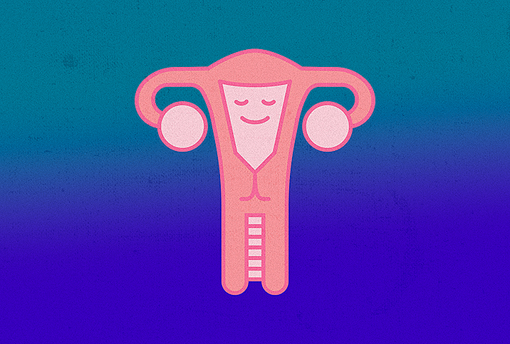
To understand adenomyosis, a lesser-known type of endometriosis, we have to understand the anatomy of the uterine lining. The wall of the uterus has three tissue layers. The endometrium is the innermost layer that makes the most direct contact with a developing fetus (or unfertilized egg).
Supporting the endometrium below are various levels of repetitive muscular patterns that power the uterus in pushing out any contents. This is known as the myometrium, which also contains its own separate levels.
'Women may need to insist on every possible exam for unexplained symptoms.'
Endometriosis is simply the presence of endometrium-like cells anywhere other than the endometrium. In cases of adenomyosis, the endometrium-like cells have penetrated the muscles supporting the uterine lining.
The tissue then proceeds to act as it would within the womb, sloughing and releasing blood into these ill-suited cavities.
Adenomyosis is rare and sometimes difficult to diagnose, but MRIs can be useful in detecting the condition. In fact, MRIs are 85 percent accurate in diagnosing the disease, according to a 2011 study published in the American Journal of Roentgenology.
The drawbacks of MRIs
MRIs have some shortcomings: they're expensive, lengthy, potentially damaging to cochlear implants or pacemakers, and may be triggering for people who have claustrophobia (although open MRIs can often be requested). Additionally, the specificity of obtaining a radiologist trained in gynecology takes effort and opportunity. However, as a second line of detection following an ultrasound or laparoscopy, the pelvic MRI can also be helpful in diagnosing particularly confounding and painful cases of endometriosis.


